
Odontoclastic Resorptive Lesions (ORLs also known as Tooth Resorptions TRs) can be a really difficult diagnosis to understand. If you have been told your pet has resorptive lesisons, you have probably also been given a lot of information to process. This page is designed to help you work understand the diagnosis and its’ implications.
What are odontoclastic resorptive lesions?
In short Odontoclastic Resorptive Lesions are painful lesions in your pets where the hardened outer layers of their teeth are being dissolved. This means the tough outer layers, comprised of enamel, dentine and outer casing is being dissolved away, and results in a very painful hole in your pet’s tooth.
We call them odontoclastic lesions, as “odontoclasts” are the specific cells in your teeth that are activated to start breaking down the teeth. When this occurs under the gumline, the resorptive tooth can often start to be replaced with bone.
You may have heard them called Feline Oral Resorptive Lesions (FORLs), Odontoclastic Resorptive Lesions (ORLs) or even just Tooth Resoptions. (TR’s) Occasionally vets will even refer to them as “an autoimmune condition that affects the teeth”. All of these refer to the same thing, and they can occur in both dogs and cats. We see it far more often in cats though, and only rarely in dogs.
What causes them?
This is the million dollar question that we would all like to know the answer to. In some cases, the cause seems to be inflammation that triggers the odontoclasts to become activated and start attacking the teeth. In other cases there seems to be something wrong with the body’s normal immune response, and the body attacks the teeth instead of the bacteria. Often this occurs below the gumline, and the tooth is gradually replaced with bone.
In most cases we don’t know what causes it, but we do know that once your pet has had one tooth resorption, it is much more likely to happen again. This is particularly true in cats.
How do you diagnose Resorptive Lesions?
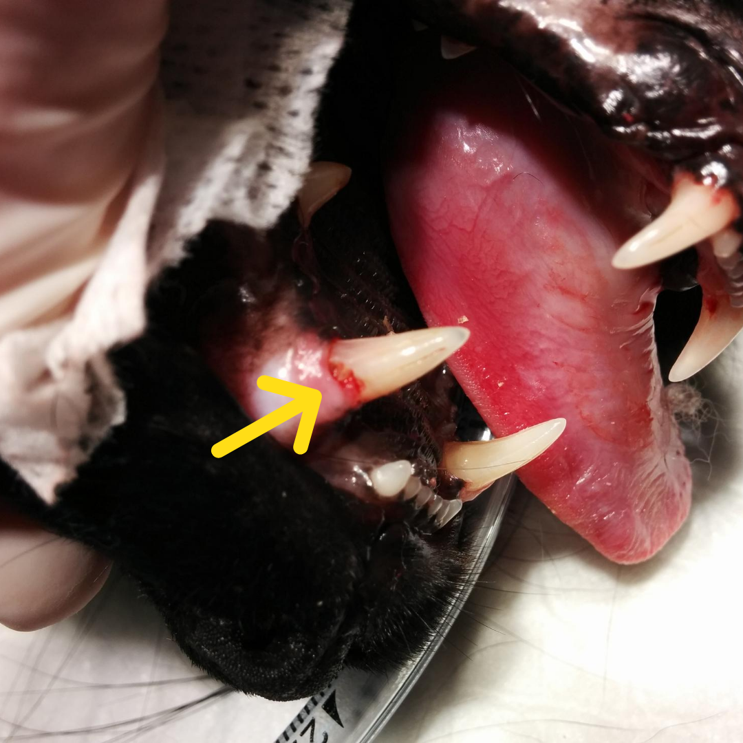
It can be difficult to diagnose without dental X-rays as animals hide their pain. Very rarely, we will have an owner notice that their pet is only eating on one side of their mouth, or has stopped eating dry food, or has started dropping more food than usual. Sometimes they notice a jaw “shudder”, or their pet drools more than usual. But most of the time, because our animals are so good at hiding pain, these lesions aren’t picked up until your pet is examined at the vet. If there is a lesion above the gum line, we can sometimes see it as a red exposed spot on your pet’s teeth. Other times we can see a “pit” in the enamel that is painful to touch and that our probes catch in.
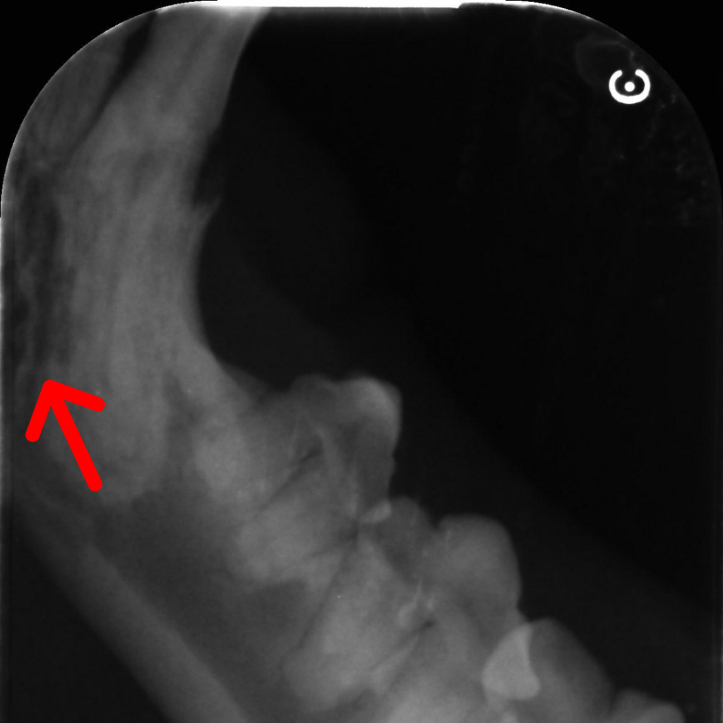
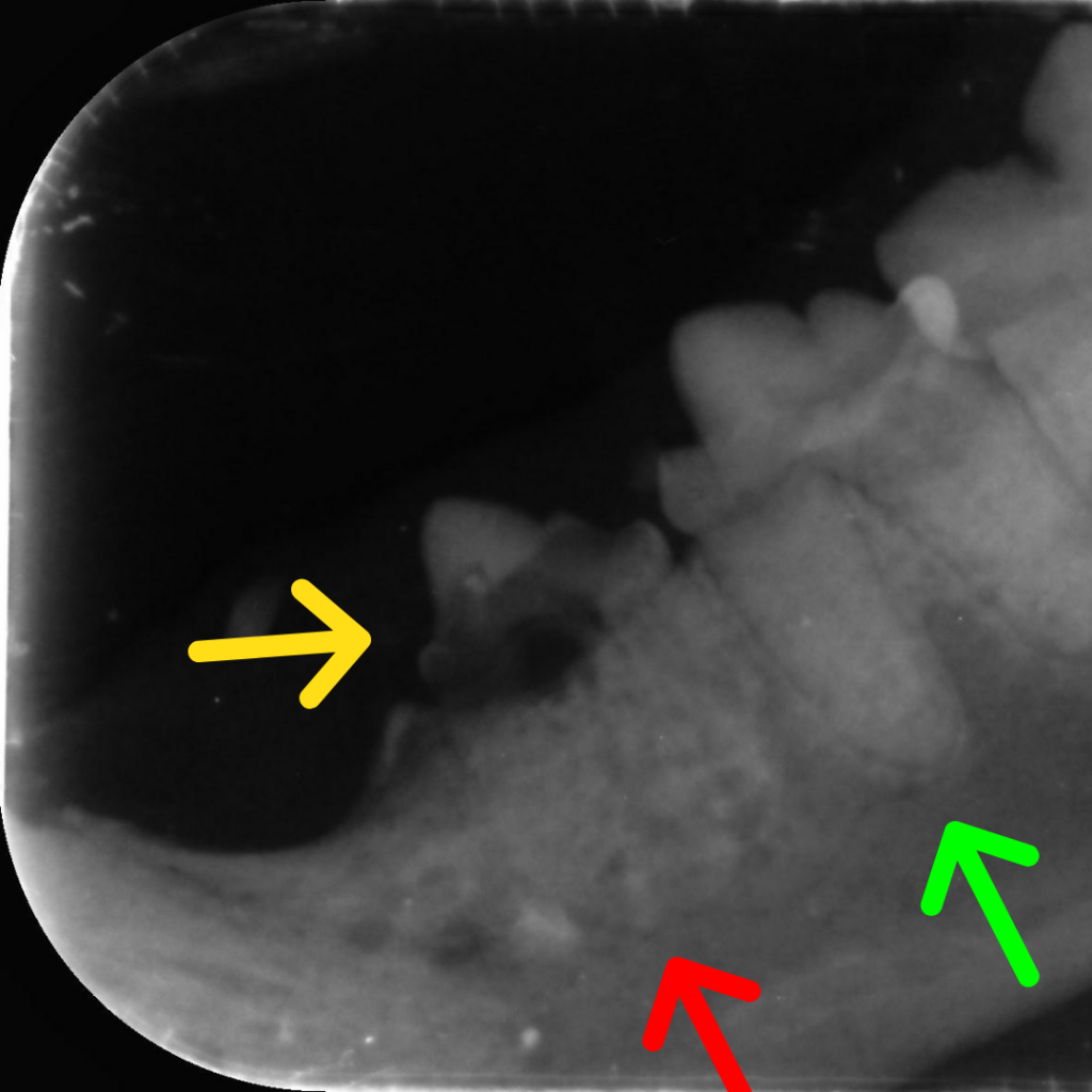
This red arrow shows that the tooth is being resorbed under the gumline. The normal periodontal ligament is missing, indicating the tooth is being replaced by bone.
Still other times, we notice gingivitis or periodontal disease and recommend a dental. It isn’t until we are taking dental X-rays of your pets mouth that we pick up that there may be resorptive lesions, as the entire lesion can occur under the gumline.
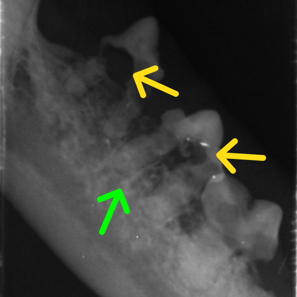
Often when the lesion is under the gum, the tooth becomes replaced with bone, and the X-rays no longer show the normal periodontal ligament around the tooth. Instead the tooth root blends into the bone around it (see Green Arrows for normal periodontal ligaments.)
Types of Resorptive Lesions
There are 3 different types of resorptive lesions.
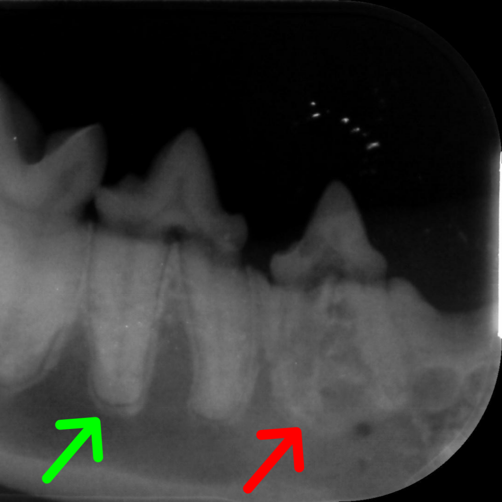
Type 1 lesions are focal, and usually above the gumline. In these cases the tooth root is usually intact, and on Xray you can see teh normal black line around the tooth root (which is the periodontal ligament). This type is most commonly associated with inflammation and gum disease.
Type 2 lesions involve the root below the gumline being resorbed and then gradually replaced with bone. The periodontal ligament is lost and the root can become difficult to distinguish from the bone around it.\
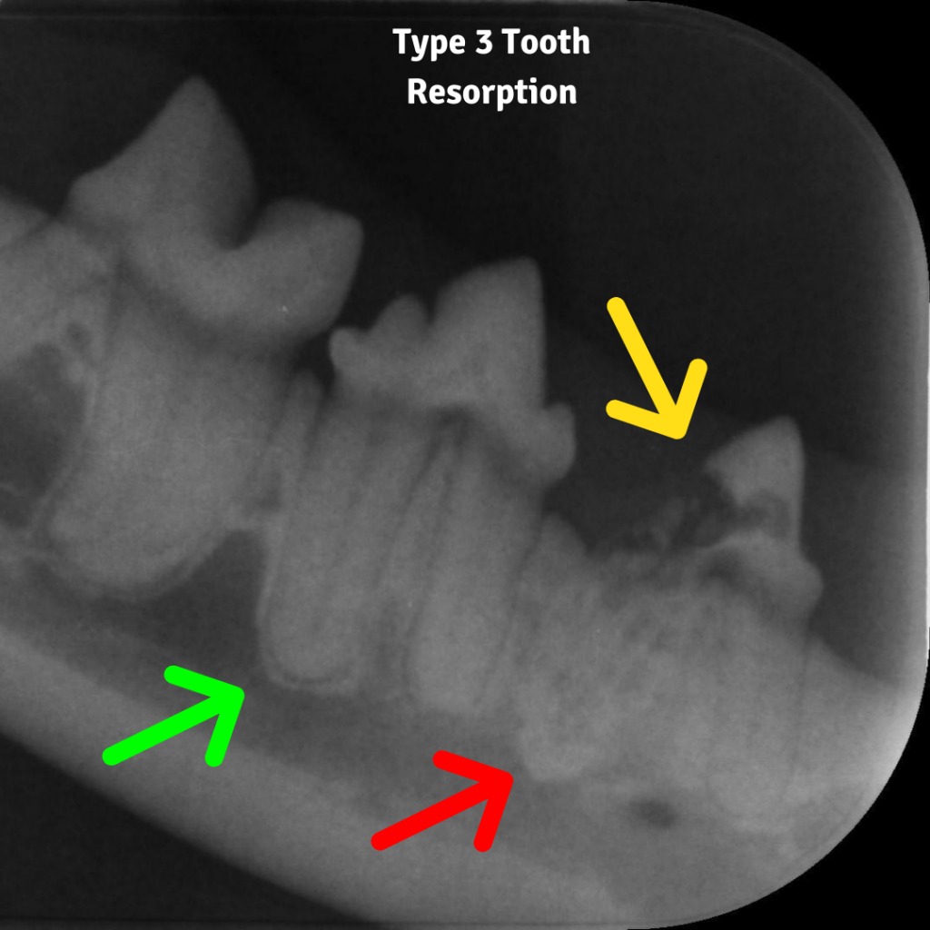
Type 3 lesions are a combination of both 1 and 2 lesions. There is focal resorptions above the gumline, but the root is also being resorbed and being replaced by bone.
How do you treat Resorptive Lesions?
This depends on the type of lesion. Type 1 lesions need to be completely removed. They are painful and cannot be removed by taking the exposed tooth away only, they need the entire root taken out as well. Type 2 and 3 can have crown amputations performed. As the root itself is already undergoing resorption and turning into bone, it often cannot be taken out and needs to be left. However in this situation, where the root is turning to bone, the root itself is not painful, and the pain resolves once the affected crown has been removed.
Does my pet really need all of their teeth taken out?
This is one of the scariest things a vet can say, but we don’t say it lightly! We almost always try to preserve as many teeth as possible.
In many cases, no, your pet does not need all of their teeth taken out, just the affected ones. But this does mean that you will need to be on the alert for recurrence. We know that once your pet has had one, they often end up with more lesions later down the track. We would usually recommend regular professional cleaning, to prevent the gum inflammation from contributing to resorptive lesions returning.
In some situations though, the best thing for your pet is to have all their teeth removed. These lesions are painful, and if your pet has advanced gum disease, or is showing other signs of on-going problems, those teeth are better out.
It is incredible how well animals do without any teeth! Cats without any teeth can often still eat dry biscuits, although some will prefer softer food. After years of pain while eating, they really do seem a lot happier and healthier without those teeth in. On those occasions when we do recommend removing all of those teeth, it is because we truly believe we will make your pet happier and more comfortable without those teeth.